Cryo-electron microscopy (cryoEM) is an ensemble of techniques allowing the observation of biological specimens in their native environment at cryogenic temperatures in EM (-180°C for liquid nitrogen stages, -269°C for He).
Low Dose Transmission Electron Microscope (TEM)
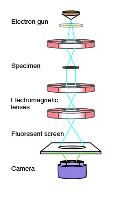
In a transmission electron microscope, an image is formed by directing a high-energy electron beam at a thin sample. A series of electromagnetic lenses collect the resulting elastically scattered electrons to form a magnified 2D image (projection) of the specimen, much like in a light microscope. The short wavelength of the electrons allows sub-nanometer resolution in these images. However a fraction of the electrons will exchange energy with the sample (inelastic scattering) and destroy it very rapidly. Therefore, for high resolution imaging the sample is only exposed to a very low electron dose (10-20 electrons Ų), and only during image acquisition.
Sample preparation
For observation in cryoEM, biological samples are preserved frozen-hydrated in their native state. This is achieved by plunging rapidly the sample reduced to a very thin liquid film into a liquid ethane bath. This operation can be computer-controlled through an automatic plunging apparatus such as the Vitrobot.


Capturing images
The resulting images are two-dimensional (2D), related to a projection of the potential in the biological object. Electron detectors can be photographic emulsions, CCD or CMOS cameras.
Data processing and Single Particle Reconstruction
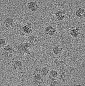
In most cases – EM image resolution is not limited by the electron wavelength or the microscope itself but from the compromise between electron dose (limited, noisy signal) and resolution to avoid electron damage. Nifty data processing is used to improve the signal quality (signal noise ratio) through averaging and reconstruction of 3D structures from the 2D data. In single particle image processing a large number of particle images are collected during one or several TEM sessions, corresponding to different angle views of identical 3D objects. A number of iterative mathematical 3D reconstruction schemes can then be applied to obtain a 3D model from these data and refine it to higher and higher resolution.
